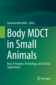
Body MDCT in Small Animals

 E’ disponibile online il primo libro di Tomografia computerizzata multidetettore nel cane e nel gatto, scritto dalla dr.ssa Giovanna Bertolini ed edito da Springer. Nato da oltre 15 anni di esperienza con questa tecnologia diagnostica e da oltre 13,000 esami eseguiti in Clinica San Marco.
RECENSIONE
BODY MDCT IN SMALL ANIMALS (Springer 2017) [AUTHOR] Bertolini, Giovanna [BIBLIOGRAPHIC DATA] ISBN: 978‐3319469027, 453 pages, hard cover. [DOODY’S NOTES] [REVIEWER’S EXPERT OPINION] Cintia R Oliveira, DVM, MS, DACVR(University of Illinois College of Veterinary Medicine)
**Description** This book on the use of thoracic and abdominal computed tomography in dogs and cats covers the most common disease processes along with pathophysiology, anatomy, and imaging description. Great emphasis is given to protocol techniques and optimization with helpful hints. Tables, graphs, and schematics make the book a pleasant read. **Purpose** The author’s purpose is to disseminate the many uses of MDCT in small animals and share her knowledge and experience in this area. The book accomplishes this by providing helpful information on imaging protocols and diagnosis. It also draws on information from human medicine to fill gaps in veterinary medicine. The book is a great addition to the imaging field. **Audience** Although intended primarily for radiologists and radiologists in training, the book also could benefit imaging technicians, as it covers imaging protocols with detailed information on angiographic studies. The author has 14 years of experience with computed tomography and is a board‐certified radiologist who has published numerous papers in the field. **Features** The author’s experience with performing and evaluating computed tomographic studies is evident throughout. The book provides comprehensive information and tailored approaches with respect to imaging techniques for each body system and sometimes for specific disease processes, so that the maximum amount of information can be obtained from each scan. Each chapter includes a concise discussion of pathophysiology and anatomy along with the most common disease process expected for the body system in question and descriptions of the computed tomographic findings. A large number of colorful images throughout the book encompass normal features as well as pathology, including schematics, multiplanar reformats, maximal intensity projection, and colorful volume‐rendered images, making the book a pleasure to read. The legends provide information about the type of study, findings, diagnosis, and patient’s clinical signs. Chapters on dual‐energy CT and cardiac CT present new insights into the multiple uses of computed tomography in veterinary medicine. Topics not explored in depth before, such as body trauma and vascular anomalies, are presented. Topics not covered in the book are head and musculoskeletal CT. This book is suitable for readers of different experience and background. It is a must‐read for radiologists as well as radiologists in training and any veterinarians or technicians who want to stay current about the multiple possibilities of multidetector computed tomography. **Assessment** This book brings an innovative and needed approach to reviewing computed tomography in veterinary medicine and is written in a clear and organized manner. The very large number of high‐quality images as well as the detailed information on angiographic studies and vascular diseases make this book unique. ‐‐‐‐‐‐‐‐‐‐‐‐‐‐‐‐‐‐‐‐‐‐‐‐‐‐‐‐‐‐‐‐‐‐‐‐‐‐‐‐‐‐‐‐‐‐‐‐‐‐‐‐‐‐‐‐‐‐‐
Weighted Numerical Score: 98 ‐ 5 Stars!


 Il Direttore Sanitario Dott. Marco Caldin